Corn vs Wart Explained: Causes, Treatment, and Easy Identification Tips
Learn the complete Corn vs Wart comparison, including causes, symptoms, treatment options, prevention strategies, and easy ways to identify the difference quickly

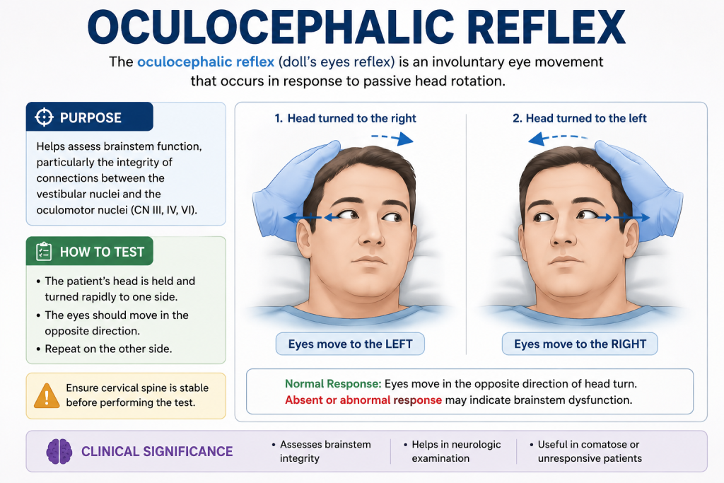
Learn about the Oculocephalic Reflex (Doll’s Eye Reflex), its mechanism, clinical significance, and 7 key facts every medical student must know in this detailed guide.
In neurology and critical care medicine, few bedside tests are as important—and as visually striking—as the Oculocephalic Reflex, also known as the Doll’s Eye Reflex. This simple yet powerful clinical test helps physicians assess brainstem function in unconscious patients.
Understanding the Oculocephalic Reflex is essential for medical students, emergency physicians, and ICU teams because it provides rapid insight into whether critical neural pathways are intact. Despite advances in imaging and diagnostics, this reflex remains a cornerstone of neurological examination.
In this comprehensive 2026 guide, we will explore the mechanism, clinical significance, testing procedure, interpretation, and key facts about the Oculocephalic Reflex that every medical student must know.
Table of contents [Show]
The Oculocephalic Reflex is a brainstem reflex that controls eye movement in response to head rotation. It is part of the vestibulo-ocular system, which stabilizes vision when the head moves.
In simple terms, when the head is turned to one side, the eyes move in the opposite direction if the brainstem is functioning properly.
This reflex is commonly tested in comatose patients and is sometimes referred to as the “Doll’s Eye Reflex” because of the doll-like movement of the eyes.
To understand the Oculocephalic Reflex, medical students must know its neural pathway:
Located in the inner ear, it detects head movement.
Transmits signals to the brainstem.
The pons and midbrain coordinate eye movement.
These nerves control eye muscles and are essential for the Oculocephalic Reflex.
The mechanism of the Oculocephalic Reflex is based on maintaining visual fixation:
If intact, the Oculocephalic Reflex confirms functional brainstem activity.
The Oculocephalic Reflex is primarily used to assess whether the brainstem is functioning in unconscious patients.
This test should NEVER be performed on awake patients, as it can cause discomfort and injury.
The Oculocephalic Reflex is strictly a clinical tool for comatose evaluation.
For a normal Oculocephalic Reflex, cranial nerves III, IV, and VI must be functional.
The Oculocephalic Reflex is one of the tests used in determining brain death protocols.
Though similar, the Oculocephalic Reflex involves head movement, while caloric testing involves cold or warm water stimulation.
An absent Oculocephalic Reflex may indicate brainstem damage or severe neurological impairment.
Incorrect testing of the Oculocephalic Reflex can lead to misinterpretation or injury in patients with cervical spine trauma.
⚠️ Important: The Oculocephalic Reflex should only be performed after cervical spine injury is ruled out.
This indicates an abnormal Oculocephalic Reflex.

The Oculocephalic Reflex is crucial in:
Helps evaluate coma depth.
Absence of Oculocephalic Reflex may support brain death criteria.
Used in head injury cases.
Helps determine brainstem involvement.
The Oculocephalic Reflex is often compared with:
Light response of pupils.
Blink response when the cornea is touched.
Induced by ear irrigation.
Each reflex, including the Oculocephalic Reflex, helps map brainstem function.
The Oculocephalic Reflex may be absent in:
Emergency physicians rely on the Oculocephalic Reflex for rapid bedside assessment because:
Medical students often make errors when assessing the Oculocephalic Reflex, such as:
Before performing the Oculocephalic Reflex, ensure:
The Oculocephalic Reflex is one of several reflexes assessed when determining brain death. Its absence alone is not sufficient but contributes to overall diagnosis.
Understanding the Oculocephalic Reflex is essential because:
Even with advanced imaging techniques like MRI and CT scans, the Oculocephalic Reflex remains a vital bedside tool due to its simplicity and reliability.
The Oculocephalic Reflex is a fundamental neurological examination tool that every medical student must understand. Despite technological advances, it remains one of the most reliable bedside assessments of brainstem integrity.
Mastering the Oculocephalic Reflex not only helps in exams but also prepares future clinicians for real-life emergency situations where quick neurological evaluation can save lives

Learn the complete Corn vs Wart comparison, including causes, symptoms, treatment options, prevention strategies, and easy ways to identify the difference quickly
Discover the best Baby Acne Treatment methods, including safe home remedies, causes, symptoms, prevention tips, and expert advice for clearing infant acne fast.
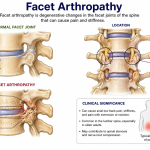
Learn everything about Facet Arthropathy, including symptoms, causes, diagnosis, and the best treatment options for managing spinal joint pain and improving mobility